Tuesday, May 16, 2006

Review artcle: Locked in Syndrome (Selectively De-efferented state)


Synonyms: Cerebromedullospinaldisconnection, Pseudocoma or de-efferented state.

Site of lesion


1. Lateral 2/3rd of cerebral peduncle bilaterally.
2. Basis pontis bilaterally sparing tegmentum.
3. Ventral aspect of medulla bilaterally sparing tegmentum.

Characteristics of the patient:


The patient is conscious, alert and awake as the tegmental ascending reticular activating system (ARAS) concerned with arousal is intact, so be careful with your bedside comments.
Blinking movements of the eyes are preserved; hence patient is able to communicate in a telegraphic pattern (“on or off” movements of eye lids).
Vertical movements are intact as it is controlled by the interstitial nucleus of cajal and the rostral part of the medial longitudinal fasciculus (MLF), which is situated in the tegmentum of the midbrain, which is spared in the locked in syndrome.
Horizontal movements of eyes are lost especially when the ventral part of the basis pontis is involved, leading to involvement of the 6th cranial nerve fascicle.
Patient may be aphonic because of the involvement of the corticobulbar fibers and motor nucleus of the lower cranial nerves especially the 7th, 9th, 10th, 12th cranial nerves.
Patient is quadriplegic due to involvement of pyramidal fibers in the lateral 2/3rd of cerebral peduncle of the midbrain or the basis pontis or the ventral aspect of the medulla.
“Fourier de prodromique” – Pathologically characteristic sudden onset of laugh at the onset of quadriparesis due to involvement of bilateral pyramidal system, producing psuedobulbar palsy like state before the onset of brainstem symptoms.


Etiology for locked in syndrome.
1.Lacunar infarcts.
2.Demeyelination(central pontine myelinolysis)
3.Haemmorhage.
4.Trauma.
5.Tumours.

Pseudo-Locked in states
1.Gullain barre syndrome
2.Acute polyneuritis
3.Myasthenia gravis
4.Poliomyelitis

References

1. Text book of neurology;Paul brazis.

2. National Institute of Neurological Diseases (NINDS) Reprot on Locked in Syndrome.

Sunday, May 14, 2006

CT corner: Schizencephaly


Schizencephaly is an uncommon disorder of neuronal migrational characterized by a cerebrospinal fluid–filled cleft, which is lined by gray matter. The cleft extends across the entire cerebral hemisphere, from the ventricular surface (ependyma) to the periphery (pial surface) of the brain.

The clefts may be unilateral or bilateral and may be closed (fused lips), as in schizencephaly type I, or separated (open lips), as in schizencephaly type II.

Presentation and outcome are variable, but patients typically present with seizures, hemiparesis, and developmental deficits. Usually, the severity of symptoms is related to the amount of brain affected by the abnormality.

References:

1.Barkovich AJ, Norman D: MR imaging of schizencephaly. AJR Am J Roentgenol 1988 Jun; 150(6): 1391-6.
2.Barkovich AJ: Schizencephaly. In: Pediatric Neuroimaging. 2nd ed. Philadelphia: Lippincott-Raven; 1996:219-25.
3.Barth PG: Schizencephaly and nonlissencephalic cortical dysplasias. AJNR Am J Neuroradiol 1992 Jan-Feb; 13(1): 104-6

Monday, May 08, 2006

Case Report: A Deadly Siren and an uncommon connective tissue disorder


Jithesh.k*, Shajith.S.*, Abhilash*, Hariprasad*, Shan** ,Geetha.P.** ,Benoy.J.Paul***.
Department of Internal medicine, Calicut Medical College.
(Published in CALFIM Journal)

Introduction
Of all the inflammatory myopathies the chance association with malignant lesions especially in the older age group is highest with dermatomyositis. However the extent of search that should be conducted for an occult neoplasm in adults depends on the clinical circumstances uncovered by the medical history and physical examination and not through an extensive blind search.
Case report
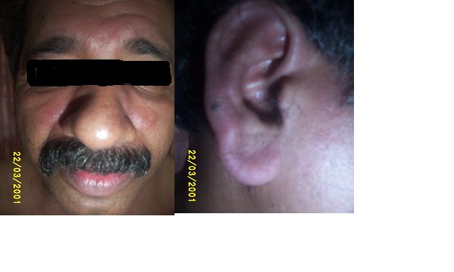
Mr........., a 48 year old previously healthy individual working in gulf countries with history of intermittent episodes of Bronchial asthma and Atopic features, presented in our outpatient department complaining of polyarthralgia involving predominantly the small joints of upper limbs and lower limbs symmetrically and generalized muscle pains for 1month and proximal muscle weakness of UL/LL and erythematous lesion of face of 3weeks duration .His physical examination showed no pallor, jaundice, cyanosis ,clubbing , edema or lymphadenopathy and pulse rate was 80/min,B.P.of 140/80 mm of Hg. His abdomen was soft with no hepatosplenomegaly or masses. Examination of chest and cardiovascular system was normal. Nervous system examination showed grade (4-) power of proximal muscles of upper and lower limbs, deep tendon reflexes were normal, with no sensory deficits, no cerebellar signs, Fundus -- normal with no overt features of peripheral neuropathy. . Locomotor system examination showed only arthralgia in the small, appendicular joints symmetrically predominantly of the upper limbs. Face showed an erythematous and edematous elevated lesions over the malar eminence on both sides involving the nasolabial folds and periocular and periorbital region, extending to the pinna on both sides.

In view of the presence of proximal weakness of the upper and lower limbs and the presence of a periorbital facial rash, the possibility of dermatomyositis was considered and we proceeded with investigations. Also a reasonable screening for occult malignancies was done to rule out secondary dermatopolimyositis.

Investigations
•Hb – 13.6 gm/dl
•TC – 7800 cells/mm3
•DC – P48 L 51 E1
•ESR – 39 mm/ I hr
•B.U. – 19 mg/dl
•S. Cr. – 0.41 mg/dl
•Na+ -- 140 meq/l
•K+ -- 4.3 meq/l
•CXR (PA) – WNL
•USG (abd) – Normal
•CPK – 7500 IU/L
•RA factor – Negative.
· Anti- ds DNA – Negative
· ANA – Negative.
Complete ANA profiling
•SS-A / Ro 52 – Positive.
•SS-B / La – Negative.
•Anti Jo -1 – Negative.
•Scl-70 – Negative.
•nRNP / Sm – Negative.
•Sm – Negative.
•CENP – Negative.
•AMA M2 – Negative
TFT
•T3 – 1.03 (0.86-2.02)
•T4 – 8.53 (5.13-14.16)
•TSH – 1.31 (0.27-4.2)
•Skin biopsy was done from the skin lesions
Showing epidermal atrophy. Epidermis shows occasional vacuolated basal cells with few scattered lymphocytes in the underlying dermis and mucinous change with an increase in the faint bluish matrix of dermis.

EMG was done to confirm with myopathic pattern of weakness, which showed spontaneous activity in the form of fibrillations and prolonged insertional activity with myopathic motor unit potentials suggestive of inflammatory myopathy.
Modified Bohan and Peter criteria were used to confirm a diagnosis of Dermatomyositis based on above clinical features and investigation reports.
Modified Bohan and Peter Criteria
.
1. Symmetrical proximal muscle weakness. (+)
2. Elevated muscle enzymes, (+)
3. Characteristic inflammatory myopathic EMG finding. (+)
4. Muscle biopsy showing evidence of inflammatory myositis.
5. Typical rash of Dermatomyositis (+)
(Three of the first Four + 5th criteria is needed for definitive diagnosis)

A reasonable screening for occult malignancies in the lung, GI system, testes and lymphoreticular system was done which was all-negative.
Patient was started on oral prednisolone at 40mg OD dose. Patient showed dramatic improvement in weakness and a decrease in CPK levels (4839 IU/l)
and skin lesion significantly decreased. Patient was kept under regular follow up to rule out any occult malignancy that may resurface in the future.

Photographs after treatment with steroids.

Discussion

Causes for Dermatomyositis
Primary

· Genetic predisposition (HLA DR3, HLA DQA1*0501).
Secondary
·
· Underlying malignancies (ovarian, breat, melanoma, colon, NHL).
·
· Autoimmune disorders (associated with MCTD or SS, rarely SLE Rhuematoid arthritis and Sjogrens syndrome).
·
· Infectious or toxic agents

· Drug-induced (implicated drugs include hydroxyurea, penicillamine, statins, quinidine, and phenylbutazone).

Treatment and follow-up of Dermatomyositis.


•Prednisone,
•Methotrexate,
• Azathioprine,
•Cyclophosphamide,
•Cyclosporin,
•Mycophenolate and
•High dose intravenous immunoglobulin.
•Diltiazem, a calcium channel blocker, may reduce calcinosis.

•Colchicine has also been reported to reduce calcinosis.

•Hydroxychloroquine may reduce the photosensitive rash.
•Avoid excessive sun exposure and use sun protection measures,

•Bed rest for those with severe inflammation of muscles,

•Physical therapy and activity to keep the muscles and joints moving.
•Raising the bed head for those with difficulty swallowing

•Most patients will require treatment throughout their lifetime,

•Completely resolves in about 20%.
· Treated DM has 5 year survival of ~95%.
•Specific Anti-Mi-1 is found in one quarter and
•Anti-Jo-1 in a few, usually those who have lung disease (80%).


•Patients who have disease affecting their heart (AV conduction abnormalities, DCM) or lungs (ILD), or who also have an underlying cancer do less well and may ultimately die from their disease.

References.

1. CALLEN JP: Dermatomyositis. Lancet 355:53, 2000

2. DALAKAS MC: Polymyositis, dermatomyositis, and inclusion-body myositis. N Engl J Med 325:1487, 1991

3. ARGOV Z et al: Various types of hereditary inclusion body myopathies map to chromosome 9p1-q1. Ann Neurol 41:548, 1997

4. ENGEL AG et al: The polymyositis and dermatomyositis syndromes, in Myology, AG Engel, C Franzini-Armstrong (eds). New York, McGraw-Hill, 1994, pp 1335-1383

5. IOANNOU Y et al: Myositis overlap syndromes. Curr Opin Rheumatol 11:468, 1999

Wednesday, May 03, 2006

Physical examnination: Vesicular breath sounds


Vesicular breath sound production

Classically the lung sounds were thought to be produced by the vibrations of the vocal cords and the proximal airways set in by the inspiratory movement of air, which is attenuated and conducted to the chest wall and the auscultating stethoscope by the normal air filled sacs called alveoli/acinus. The normal breath sounds were initially thought to be produced in the terminal air filled alveoli, and was called Vesicular sounds (Vesicle = small vessels). Present view is that the terminal airways including acinus are silent due to the laminar flow and low velocity of airflow,and breath sounds are solely producd by the vibrations of the proximal airways. As the vibrations of taut vocal cord can produce harmonics of both high and low pitches, and the normal vesicular breath sound is low pitched with a frequency of 16-200HZ, further supports the earlier proposed mechanism of attenuation and conduction of sounds by the terminal air spaces.

Definition of vesicular breath sounds

ATS definition of vesicular breath sound -- VBS is a low pitched rustling sound with a high intensity inspiratory phase which is continuous with a less intense and shorter expiratory phase, which normally extends to less than one third of inspiratory phase.

Now look at the first part of the diagram for the possible site of origin of each component of vesicular breath sound.

Look at the second part of the diagram for diagrammatic representation of normal vesicular breath sound (inverted and mirror image for better understanding).

Causes for decreased intensity of normal vescular breath sound.
1. Pleural effusion (Separation of conducting units from chest wall)
2. Pleural thickening (-do-)
3. Collapsed lung with occluded bronchus (Loss of conducting pathways)
4. Emphysema (Loss of laminar flow and low velocity airflow due to distended acinus)
5. Bronchial asthma (occlusion of conducting airways)
6. Thick chest wall (eg;Obesity)(separation of steth from the area of activity” lung”).

References

1. Kraman,SS: Vesicular(normal) lung sounds:How are they made,where do they come from,and what do they man?; Semi. In Resp Med 6:183,1985.
2. Forgacs,P: The functional basis of pulmonary sounds, Chest 73:399,1978.

Tuesday, May 02, 2006

Gastroenterology:Traube's space


Surface Markings

1. Draw two vertical lines one passing through the 6th rib in the midclavicular line and the next passing through the 9th rib in midaxillary lines.

2. Now draw a smooth curving line with convexity upwards ftom the sixth rib in midclavicular line to 9th rib in midaxillary line.

3.Draw another straight line passing through the costal margin from 6th rib to 9th rib.

All these boundaries enclose a near semilunar space called Traubes space.

Anatomical boundaries are:

1. Right : Lateral margin of left lobe of liver.
2. Left : Spleen.
3. Superior : Resonance of lung.
4. Inferior : Costal margin.

Contents

1. Fundus of stomach (Hence percussion of Traubes area normally gives Tympanitic resonance).
2. Costo-phrenic recess of left pleura devoid of lungs.

Causes of obliteration of Traubes space:
1. Full stomach.
2. Left sided Pleural effusion.
3.Splenomegaly.
4. Enlargment ofleft lobe of liver due to any etiology.
5. Dextrocardia.
6. Proloiferative growth in fundus of stomach.

Note: A left lung mass lesion/consolidation alone never produces impairment as lung is not extending to traube's space.